File:Bronchiolar epithelium 3 - SEM.jpg
Jump to navigation
Jump to search

Size of this preview: 585 × 599 pixels. Other resolutions: 234 × 240 pixels | 469 × 480 pixels | 750 × 768 pixels | 1,024 × 1,049 pixels.
Original file (1,024 × 1,049 pixels, file size: 375 KB, MIME type: image/jpeg)
Captions
Captions
Add a one-line explanation of what this file represents
Summary[edit]
| Description |
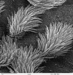
Scanning electron microscope image of lung trachea epithelium. There are both ciliated and non-ciliated cells in this epithelium. Note the difference in size between the cilia and the microvilli (on the non-ciliated cell surface). Zeiss DSM 962 SEM |
| Source | |
| Author | Charles Daghlian |
| Permission (Reusing this file) |
PD |
Licensing[edit]
| This work has been released into the public domain by its author, Charles Daghlian. This applies worldwide. In some countries this may not be legally possible; if so: Charles Daghlian grants anyone the right to use this work for any purpose, without any conditions, unless such conditions are required by law.
|
File history
Click on a date/time to view the file as it appeared at that time.
| Date/Time | Thumbnail | Dimensions | User | Comment | |
|---|---|---|---|---|---|
| current | 14:16, 7 October 2006 | | 1,024 × 1,049 (375 KB) | Patho (talk | contribs) | {{Information |Description=Scanning electron microscope image of lung trachea epithelium. There are both ciliated and on-ciliated cells in this epithelium. Note the difference in size between the cilia and the microvilli(on non-ciliated cell surface) Zei |
You cannot overwrite this file.
File usage on Commons
There are no pages that use this file.
File usage on other wikis
The following other wikis use this file:
- Usage on ar.wikipedia.org
- Usage on ast.wikipedia.org
- Usage on bs.wikipedia.org
- Usage on ca.wikipedia.org
- Usage on cs.wikipedia.org
- Usage on da.wikipedia.org
- Usage on de.wikipedia.org
- Usage on de.wikibooks.org
- Usage on en.wikipedia.org
- Usage on es.wikipedia.org
- Usage on eu.wikipedia.org
- Usage on fa.wikipedia.org
- Usage on fr.wikipedia.org
- Usage on gl.wikipedia.org
- Usage on he.wikipedia.org
- Usage on he.wiktionary.org
- Usage on hi.wikipedia.org
- Usage on id.wikipedia.org
- Usage on jv.wikipedia.org
- Usage on kk.wikipedia.org
- Usage on lt.wikipedia.org
- Usage on lv.wikipedia.org
- Usage on ms.wikipedia.org
- Usage on nl.wikipedia.org
- Usage on nn.wikipedia.org
- Usage on no.wikipedia.org
- Usage on pl.wikipedia.org
- Usage on pl.wiktionary.org
- Usage on pt.wikipedia.org
- Usage on ru.wikipedia.org
- Usage on ru.wiktionary.org
- Usage on sh.wikipedia.org
View more global usage of this file.